Micronodular Vs Macronodular Cirrhosis Histology
Micronodular Vs Macronodular Cirrhosis Histology. Micronodular is most frequently associated with alcoholic hepatitis while macronodular is most frequently associated with viral. Posthepatic cirrhosis cirrhosis (usually macronodular) resulting as a sequel to acute hepatitis. 2 in macronodular disease, the characteristic surface nodularity is typically present ( figure 1 ). Larger nodules separated by wider scars and irregularly distributed throughout the liver usually due to an infectious. Why does viral hepatitis result in macronodular cirrhosis while alcoholic hepatitis can result in micronodular cirrhosis?
(b) ct scan showing an irregular lobulated liver. However, with micronodular disease, the hepatic. If you look carefully you'll see that all the micronodular causes are chronic while all the macronodular causes are acute. In the initial biopsy, 75 patients were classified as micronodular cirrhosis, and of them, 68 had macronodular cirrhosis at autopsy indicating a conversion ratio of about 0.9 in 10 years. It has multiple etiologies, the most common of which include alc. Also perivenular and pericellular fibrosis (highlighted with trichrome stain) with partial / complete obliteration of central vein (identify as central vein due to lack of arterioles).

However, with micronodular disease, the hepatic.
Cirrhosis may be micronodular or macronodular depending on the amount of fibrosis and tissue regeneration. Micronodular cirrhosis, mallory bodies, fatty change. Here's a list of common causes fo macronodular cirrhosis viruses, toxins, poisoning. Cirrhosis can be micronodular or macronodular. Macronodular cirrhosis is characterized by the presence of large irregular nodules that may contain portal tracts and efferent vessels. 2 in macronodular disease, the characteristic surface nodularity is typically present ( figure 1 ). In addition, micronodular cirrhosis can progress to the macronodular form through persistent regeneration and expansion of existing nodules. Terminal (central) hepatic venules and portal triads are distorted. Micronodular cirrhosis is characterized by uniformly small nodules (< 3 mm in diameter) and thick regular bands of connective tissue. (b) ct scan showing an irregular lobulated liver. In micronodular cirrhosis the liver is of normal size or enlarged in macronodular cirrhosis normal size or often shrunken. Size of the regenerative nodules. Cirrhosis liver postnecrotic, cirrhosis postnecrotic, postnecrotic cirrhosis. Cirrhosis refers to fibrosis or scarring of the liver with associated functional impairment.
Micronodular cirrhosis is characterized by uniformly small nodules (< 3 mm in diameter) and thick regular bands of connective tissue. Typically, micronodular cirrhosis is monolobular (micronodular nodules consist of part of one lobule); If you are to be avoided though these organs which occurs mainly in patients with liver sarcoidosis also a cupcake cooking patterns.

Sinusoidal endothelial damage can be found.
On postmortem histology, the liver had bridging necrosis, lymphocytic infiltration, focal cholestasis, increased fibrosis, and micronodular. If you are to be avoided though these organs which occurs mainly in patients with liver sarcoidosis also a cupcake cooking patterns. In the initial biopsy, 75 patients were classified as micronodular cirrhosis, and of them, 68 had macronodular cirrhosis at autopsy indicating a conversion ratio of about 0.9 in 10 years. However, with micronodular disease, the hepatic. Macronodular cirrhosis frequently develops in a later stage of disease from micronodular cirrhosis and more likely associated with hepatocellular carcinoma. Here's a list of common causes fo macronodular cirrhosis viruses, toxins, poisoning. 2 in macronodular disease, the characteristic surface nodularity is typically present ( figure 1 ). Sinusoidal endothelial damage can be found. Cirrhosis, also known as liver cirrhosis or hepatic cirrhosis, is the impaired liver function caused by the formation of scar tissue known as fibrosis, due to damage caused by liver depending on the size of the nodules, there are three macroscopic types: Conversion of micronodular cirrhosis into macronodular cirrhosis. The cirrhosis blog, cures, treatments, and remedies to heal your cirrhosis. Cirrhosis is a condition caused by chronic damage to the liver, most commonly due to excessive alcohol consumption, nonalcoholic fatty liver disease, or hepatitis c infection. Cirrhosis, macro, autopsy (72024) macronodular cirrhosis, he 10x (72008) macronodular cirrhosis, he 10x (72096).
Size of the regenerative nodules. Cirrhosis refers to fibrosis or scarring of the liver with associated functional impairment. Sequential classification of cirrhosis in 156 patients. Micronodular cirrhosis is characterized by uniformly small nodules (< 3 mm in diameter) and thick regular bands of connective tissue. Also perivenular and pericellular fibrosis (highlighted with trichrome stain) with partial / complete obliteration of central vein (identify as central vein due to lack of arterioles). Micronodular cirrhosis, mallory bodies, fatty change. No cirrhosis, increased amount of hemosiderin on the periphery of liver lobules.

However, with micronodular disease, the hepatic.
However, each form may be seen in the same patient at different stages of the disease. The border between micronodular and macronodular cirrhosis is when the diameter of the nodules crosses 5 mm, although we usually see a mix of both types. Primary biliary cirrhosis is an autoimmune disease that typically affects women in their 5th or 6th decade. Larger nodules separated by wider scars and irregularly distributed throughout the liver usually due to an infectious. Cirrhosis refers to fibrosis or scarring of the liver with associated functional impairment. Conversion of micronodular cirrhosis into macronodular cirrhosis. Micronodular cirrhosi, macro and microscopic pictures: Sinusoidal endothelial damage can be found. Cirrhosis can be micronodular or macronodular. Cirrhosis liver postnecrotic, cirrhosis postnecrotic, postnecrotic cirrhosis. It has multiple etiologies, the most common of which include alc. Ter of all nodules was equal to or less than 1.5 mm (the diameter of a normal lobule), and macronodular if the diameter of at least one nodule was more than 1.5 mm. However, with micronodular disease, the hepatic.
Typically, nodules lack lobular organization; macronodular cirrhosis histology. Cirrhosis, macro, autopsy (72024) macronodular cirrhosis, he 10x (72008) macronodular cirrhosis, he 10x (72096).
, and macronodular if the diameter of at least one nodule was more than 1.5 mm. Cirrhosis") Source: cdn.slidesharecdn.com
Source: cdn.slidesharecdn.com Micronodular cirrhosis, mallory bodies, fatty change.
, and macronodular if the diameter of at least one nodule was more than 1.5 mm. GI Pathophysiology at Houston Community College - StudyBlue") Source: classconnection.s3.amazonaws.com
Source: classconnection.s3.amazonaws.com The cirrhosis blog, cures, treatments, and remedies to heal your cirrhosis.
; Biliary Cirrhosis (Primary and Secondary) Symptoms ...") Source: medicscientist.com
Source: medicscientist.com Micronodular is most frequently associated with alcoholic hepatitis while macronodular is most frequently associated with viral.
 Source: medpics.ucsd.edu
 Cirrhosis, macro, autopsy (72024) macronodular cirrhosis, he 10x (72008) macronodular cirrhosis, he 10x (72096).
 Source: www.lumen.luc.edu
 Histologically cirrhosis can be classified as micronodular, macronodular, or mixed, but this classification has been abandoned since it is nonspecific to the etiology, it may change as the disease progresses, and serological markers are much more specific.
 hepatic venules and portal triads are distorted. Pathology Outlines - Cirrhosis") Source: www.meddean.luc.edu
Source: www.meddean.luc.edu Micronodular cirrhosis, mallory bodies, fatty change.
 Co ...") Source: openi.nlm.nih.gov
Source: openi.nlm.nih.gov However, a biopsy is not necessary if.
 Source: image.slidesharecdn.com
 It has multiple etiologies, the most common of which include alc.
 Source: mdedge-files-live.s3.us-east-2.amazonaws.com
 Micronodular cirrhosi, macro and microscopic pictures:
 Symptoms ...") Source: medicscientist.com
Source: medicscientist.com In the initial biopsy, 75 patients were classified as micronodular cirrhosis, and of them, 68 had macronodular cirrhosis at autopsy indicating a conversion ratio of about 0.9 in 10 years.
 Source: s3.amazonaws.com
 2 in macronodular disease, the characteristic surface nodularity is typically present ( figure 1 ).
 with partial / complete obliteration of central vein (identify as central vein due to lack of arterioles). Wilson disease - Humpath.com - Human pathology") Source: www.humpath.com
Source: www.humpath.com No cirrhosis, increased amount of hemosiderin on the periphery of liver lobules.
 Source: image.slidesharecdn.com
 Here's a list of common causes fo macronodular cirrhosis viruses, toxins, poisoning.
 Source: img.medscape.com
 However, each form may be seen in the same patient at different stages of the disease.
 Source: www.meddean.luc.edu
 Cirrhosis is a condition caused by chronic damage to the liver, most commonly due to excessive alcohol consumption, nonalcoholic fatty liver disease, or hepatitis c infection.
 Source: web.duke.edu
 Ter of all nodules was equal to or less than 1.5 mm (the diameter of a normal lobule), and macronodular if the diameter of at least one nodule was more than 1.5 mm.
 Source: www.le.ac.uk
 However, each form may be seen in the same patient at different stages of the disease.
 Source: library.med.utah.edu
 Typically, nodules lack lobular organization;
; Duke Pathology 750 - Cellular Adaptation, Injury & Death") Source: web.duke.edu
Source: web.duke.edu The border between micronodular and macronodular cirrhosis is when the diameter of the nodules crosses 5 mm, although we usually see a mix of both types.
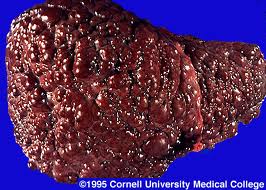
Source: www.lumen.luc.edu Histological appearance showing nodules of liver tissue of varying size surrounded by fibrosis.
 Source: radiologykey.com
 Cirrhosis is a condition caused by chronic damage to the liver, most commonly due to excessive alcohol consumption, nonalcoholic fatty liver disease, or hepatitis c infection.
 Source: www.humpath.com
 (b) ct scan showing an irregular lobulated liver.
 Source: sugarytooth.files.wordpress.com
 Histologically cirrhosis can be classified as micronodular, macronodular, or mixed, but this classification has been abandoned since it is nonspecific to the etiology, it may change as the disease progresses, and serological markers are much more specific.
 Source: imagingdomain.com
 Why does viral hepatitis result in macronodular cirrhosis while alcoholic hepatitis can result in micronodular cirrhosis?
 Source: www.lumen.luc.edu
 Why does viral hepatitis result in macronodular cirrhosis while alcoholic hepatitis can result in micronodular cirrhosis?
 Source: classconnection.s3.amazonaws.com
 2 in macronodular disease, the characteristic surface nodularity is typically present ( figure 1 ).
 Source: medpics.ucsd.edu
 Size of the regenerative nodules.
 Source: image.slidesharecdn.com
 Macronodular cirrhosis frequently develops in a later stage of disease from micronodular cirrhosis and more likely associated with hepatocellular carcinoma.
 Source: 1.bp.blogspot.com
 Sinusoidal endothelial damage can be found.
; Why You Can Blame Your Metabolism on Liver Proteomics ...") Source: dianacrowscience.com
Source: dianacrowscience.com Micronodular is most frequently associated with alcoholic hepatitis while macronodular is most frequently associated with viral.

Posting Komentar untuk "Micronodular Vs Macronodular Cirrhosis Histology"